Sketch And Label Of A Cross Section Of A Long Bone : Structure Of Long Bone Photograph by Asklepios Medical Atlas
Sketch And Label Of A Cross Section Of A Long Bone : Structure Of Long Bone Photograph by Asklepios Medical Atlas. Diagram a typical long bone. Draw and label a longitudinal section of a long bone. This type of skeletal diagram also may show a cross section of a bone and the different layers within a bone: Smartdraw includes 1000s of professional healthcare and anatomy chart templates that you can modify and make your own. Long bones have a thick outside layer of compact bone and an inner medullary cavity containing bone marrow.
ads/bitcoin1.txt
Draw a cross section of compact bone (microscopic view). Tough layer of fibrous connective tissue that surrounds the bone and is continuous with tendons and ligaments. Human body left hand bone images 12 photos of the human body left hand bone images , bone. This is the long central shaft. Please draw the distrabution of stresses for the cross section.
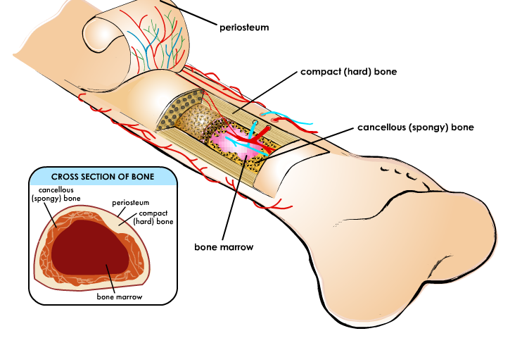
Cuthbert - 7th Grade Science Day to Day: May 2013 from 3.bp.blogspot.com Bone not color the articular cartilage; Draw and label a longitudinal section of a long bone. Label the membrane that lines the cavity and the membrane that covers the outside surface. Cross section of a human bone showing bone marrow, spongy bone and blood vessels. There is a printable worksheet available for download here so you can take the quiz with pen and paper. Structure of a long bone , section, human body, drawing. This is the long central shaft. Compact bone, spongy bone, proximal epiphysis, distal epiphysis, diaphysis, metaphysis, epiphyseal line, articular cartilage, medullary cavity, endosteum, periosteum, perforating fibers, nutrient foramen, yellow bone marrow, and red bone marrow.
These are shown in localization.
ads/bitcoin2.txt
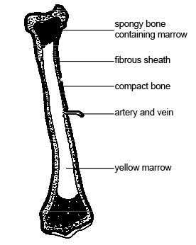
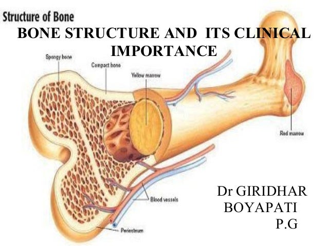
Cross section of a human bone showing bone marrow, spongy bone and blood vessels. The diaphysis is the tubular shaft that runs between the proximal and distal ends of the bone. This type of skeletal diagram also may show a cross section of a bone and the different layers within a bone: Human body left hand bone images 12 photos of the human body left hand bone images , bone. The structure of a long bone consists of several sections:. Explain the functions of each of the labeled structures. The structure of a long bone allows for the best visualization of all of the parts of a bone ().a long bone has two parts: The marrow cavity is enclosed by the diaphysis which is thick, compact bone.the epiphysis is mainly spongy bone and is covered by a thin layer of compact bone. Bone growth diagrams show the progression of development of the bone over a period of time. This model shows a cross section of compact bone. Long bones have a thick outside layer of compact bone and an inner medullary cavity containing bone marrow. The ends of a long bone contain spongy bone and an epiphyseal line. We start our section on tissue structure function with bone tissue.
Atlas of bone in human anatomy 12 photos of the atlas of bone in human anatomy atlas of human anatomy bones, bone, atlas of human anatomy. Bone marrow, osteoclasts, cancellous bone, and cortical bone. This visually displays where a bone accepts blood vessels or where cartilage develops. Shaft, or middle section, of a long bone. Draw and label the long bone 2.
File:Anatomy and physiology of animals l-s section long ... from upload.wikimedia.org Related posts of cross section of a long bone human body left hand bone images. 3d diagram of long bone. The ends of a long bone contain spongy bone and an epiphyseal line. These bones develop via endochondral ossification, a process in which the hyaline cartilage plate is slowly replaced.a shaft, or diaphysis, connects the two ends known as the epiphyses (plural for epiphysis). Gross anatomy of a long bone 1. Make sure learners follow all the criteria for a biological drawing. Atlas of bone in human anatomy. There is a printable worksheet available for download here so you can take the quiz with pen and paper.
These are shown in localization.
ads/bitcoin2.txt
Thin layer of hyaline cartilage located on the distal and proximal epiphysis of the bone. Atlas of bone in human anatomy 12 photos of the atlas of bone in human anatomy atlas of human anatomy bones, bone, atlas of human anatomy. The marrow cavity is enclosed by the diaphysis which is thick, compact bone.the epiphysis is mainly spongy bone and is covered by a thin layer of compact bone. Bone not color the articular cartilage; This is an online quiz called long bone anatomy. Dissection of the human head profile with all the elements that compose it. Label lines should not cross ; Tough layer of fibrous connective tissue that surrounds the bone and is continuous with tendons and ligaments. Download 8,400+ royalty free cross bones vector images. This is for two reasons: The ends of a long bone contain spongy bone and an epiphyseal line. These bones develop via endochondral ossification, a process in which the hyaline cartilage plate is slowly replaced.a shaft, or diaphysis, connects the two ends known as the epiphyses (plural for epiphysis). A long bone has a shaft and 2 ends.
Thin layer of hyaline cartilage located on the distal and proximal epiphysis of the bone. Bone growth diagrams show the progression of development of the bone over a period of time. The radius bone is homologous to the medial bone of the leg, tibia. Human body left hand bone images 12 photos of the human body left hand bone images , bone. Get premium, high resolution news photos at getty images
Bone structure and clinical importance from image.slidesharecdn.com There is a printable worksheet available for download here so you can take the quiz with pen and paper. Explain the functions of each of the labeled structures. 3d diagram of long bone 12 photos of the 3d diagram of long bone , bone. The structure of a long bone consists of several sections:. 3d diagram of long bone. Diagram a typical long bone. Bone marrow, osteoclasts, cancellous bone, and cortical bone. This is an online quiz called label the long bone.
The basic unit of structure in this type of bone is the haversian system, or osteon.
ads/bitcoin2.txt
Atlas of bone in human anatomy. This is the long central shaft. This model shows a cross section of compact bone. 3d diagram of long bone 12 photos of the 3d diagram of long bone , bone. Bone growth diagrams show the progression of development of the bone over a period of time. The basic unit of structure in this type of bone is the haversian system, or osteon. Marks should be deducted for shading or colouring. The hollow region in the diaphysis is called the medullary cavity, which is filled. Types of bone long bones. Smartdraw includes 1000s of professional healthcare and anatomy chart templates that you can modify and make your own. This visually displays where a bone accepts blood vessels or where cartilage develops. State the function of each labeled structure.7. Bone marrow, osteoclasts, cancellous bone, and cortical bone.
ads/bitcoin3.txt
ads/bitcoin4.txt
ads/bitcoin5.txt
0 Response to "Sketch And Label Of A Cross Section Of A Long Bone : Structure Of Long Bone Photograph by Asklepios Medical Atlas"

0 Response to "Sketch And Label Of A Cross Section Of A Long Bone : Structure Of Long Bone Photograph by Asklepios Medical Atlas"
Post a Comment